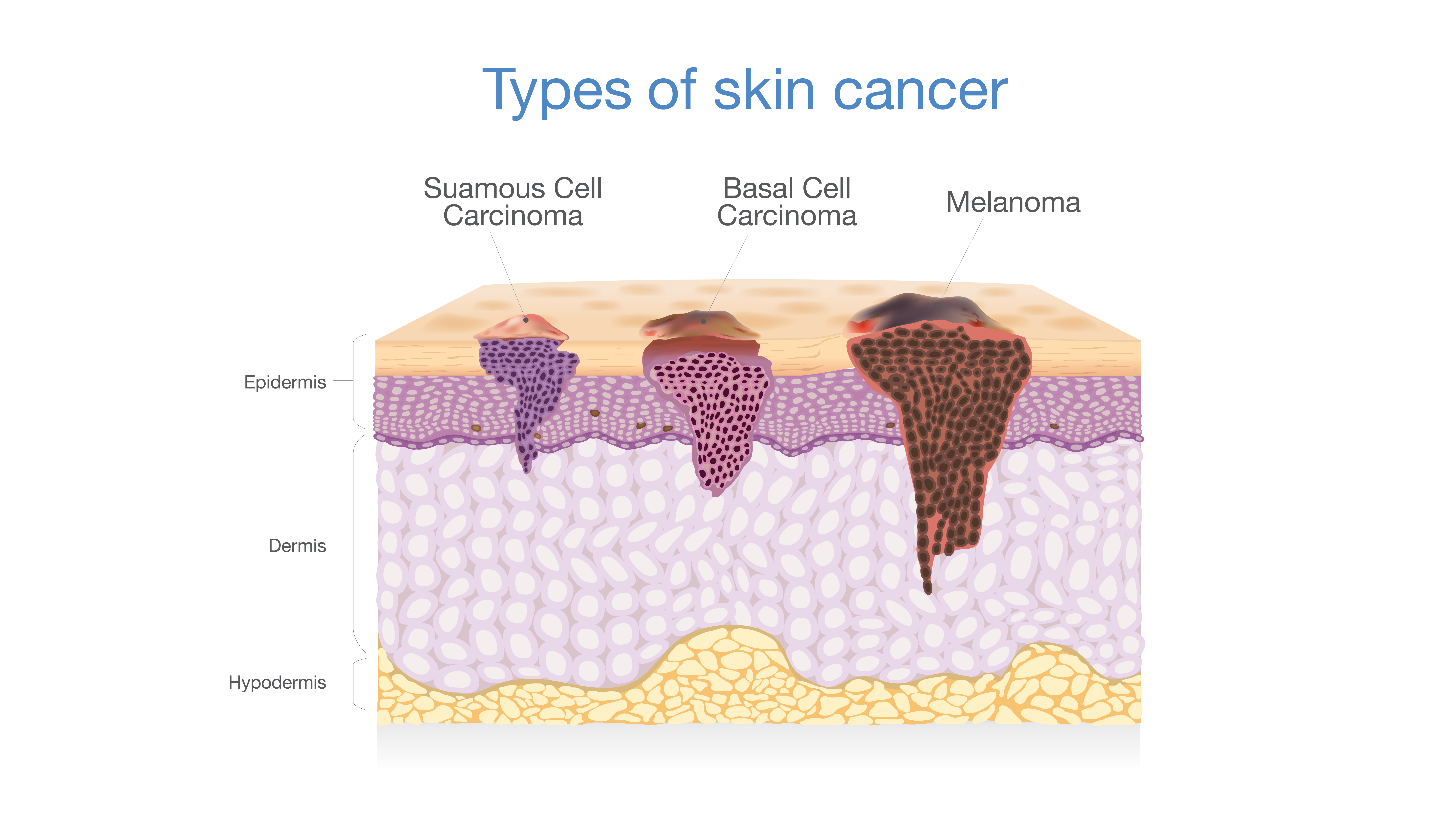
Basal Cell Carcinoma
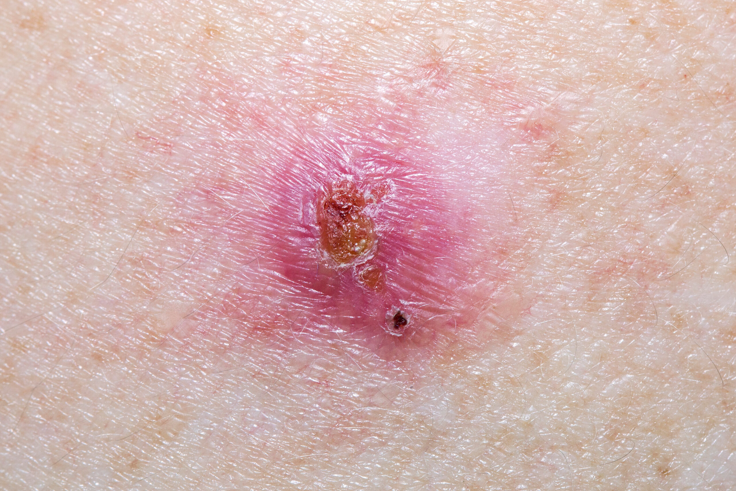
Basal Cell Carcinoma is the most prevalent form of skin cancer. Largely the result of exposure to the sun, it appears as a blemish that refuses to heal. These lesions usually appear most commonly on sun exposed skin.
What does it look like?
-
- Basal Cell Carcinomas can have many different features which is why it is important to have a qualified physician or dermatologist to assess a concerning spot.
- The most common warning signs are an area may be prone to recurrent bleeding, crusting, and the spot just won’t heal.
- Other features include:
- Slight raised, firm reddish bump
- Pearly or shiny texture
- Red scaly patch
- A sore or pimple-like growth that will not heal.
Who's at risk?
Basal Cell carcinoma is most prevalent in patients of fair skin types. Patients that have blonde or red hair, and skin the burns easily and never tans are most at risk. Age is also a factor in that the older a patient is the more sun exposure their skin has. As well, being immunocompromised from an organ transplant increases the risk of developing a Basal Cell Carcinoma.
Treatment
- Prevention is key! Covering or using sunscreen regularly can reduce the risk of developing a Basal Cell Carcinoma.
- If left untreated, Basal Cell Carcinoma can affect the surrounding tissue, nerves, and blood vessels. Fortunately, Basal Cell Carcinoma rarely ever spreads to lymph nodes or other organs.
- A skin biopsy to the area of concern is completed to determine if the lesion is truly diagnosed as a Basal Cell Carcinoma. Once the diagnosis has been confirmed then the physician or dermatologist will consider the next step in treating the Basal Cell Carcinoma.
- Treatment options include further excision via a deep biopsy, Wide Local Excision or Mohs Micrographic Surgery.
Squamous Cell Carcinoma
Squamous Cell Carcinoma is the second most common form of skin cancer, is also caused by prolonged exposure to the sun. However, Squamous Cell Carcinoma can appear on any body part, even areas that are not sun exposed.
What does it look like?
-
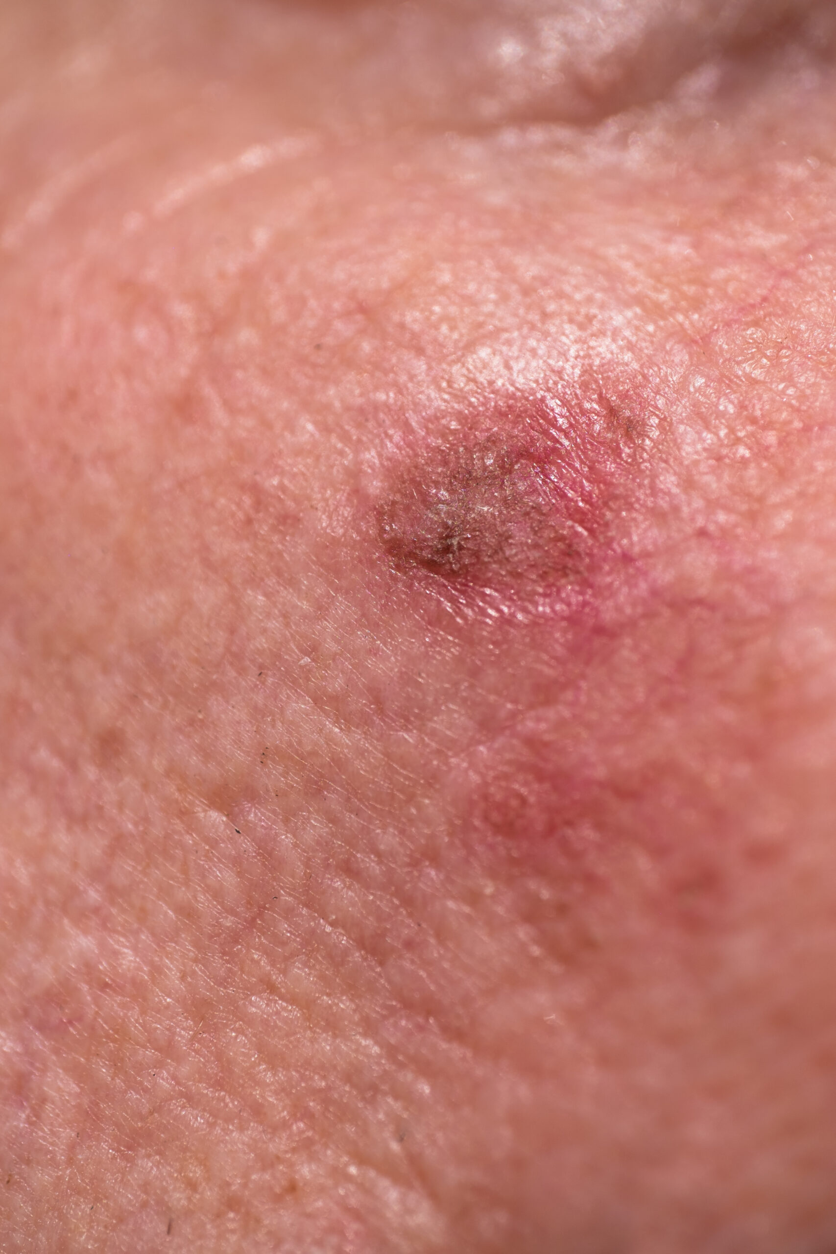
- Squamous Cell Carcinomas can start out by looking like a thick, dry, scaly bump or wartlike patch.
- The most common warning signs are an area may be prone to recurrent bleeding, crusting, and the spot just won’t heal.
Treatment
- Prevention is key! Covering or using sunscreen regularly can reduce the risk of developing a Squamous Cell Carcinoma.
- A small number of SCC can be aggressive and can spread to the lymph nodes and other internal organs if left untreated. This can lead to several other complications.
- A skin biopsy to the area of concern is completed to determine if the lesion is truly diagnosed as a Squamous Cell Carcinoma. Once the diagnosis has been confirmed then the physician or dermatologist will consider the next step in treating the Squamous Cell Carcinoma.
- Treatment options include further excision via a deep biopsy, Wide Local Excision or Mohs Micrographic Surgery.
Who's at risk?
- Squamous Cell Carcinomas are seen more commonly in patient with fair skin types and that have a history of sun exposure or exposure to tanning beds. As well, being immunocompromised from an organ transplant increases the risk of developing a Squamous Cell Carcinoma.
- Lesions found on the lip or ears are considered a high-risk area, due to the high prevalence of spreading to surround tissues and lymph nodes.
Melanoma
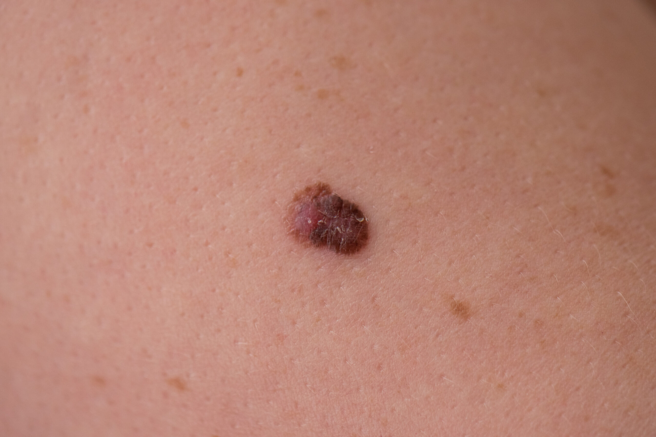
Melanoma often appears as a non-uniform dark patch or a freckle of unusual colour, this is a less common type of skin cancer but is the most serious.
What does it look like?
-
- Melanoma can develop over weeks, months, or years. It can start out as a new mole on the skin, or even develop in an existing mole.
- Melanomas are dark in color (black or brown) but can also have other colors such as blue, red, purple, or even grey.
- Melanomas also develop a peculiar shape, that would be different then the rest of a patient regular moles. This is called the “Ugly Duckling Sign” where a mole does not look like the rest of a patient moles.
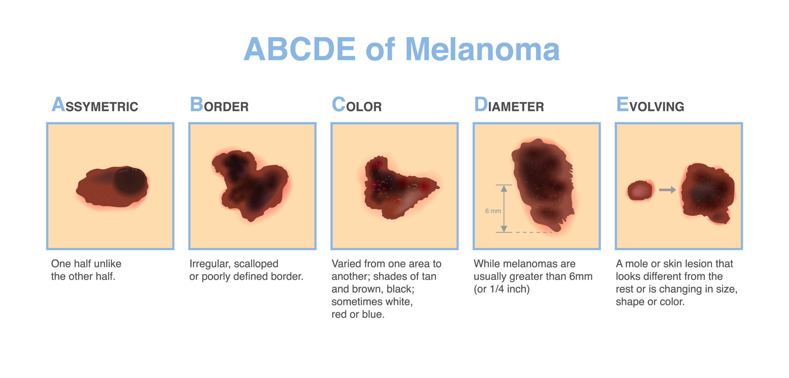
- An easy way to monitor your moles for Melanoma is as follows:
- A – Asymmetry – where the shape of one side is different than the other (does not mirror image itself)
- B – Border – are the edges of the mole clearly visible, or are the hard to determine (“fuzzy”)
- C- Colour – is the color of the mole uniform, or does it have multiple colors
- D – Diameter – has the mole grown large recently
- E – Evolution – have you notice the mole changing, does it get itchy at time, is it tender or occasionally bleed on its own
Who's at risk?
- Melanoma is more likely to develop in patients that have:
- Fair skin that burns easily when exposed to sun
- Naturally has several moles across the whole body
- A close family history of melanoma
- Excessive exposure to UV or tanning beds
- Although rare, melanoma can also develop in patient of darker skin types.
- It is important to be seen by a physician or dermatologist if you are concerned about a changing mole.
Treatment
- Fortunately, Melanomas can be cured through surgery if diagnosed and treated at an early stage.
- Just like many other types of cancer, Melanomas are graded or staged from 0 to 4, with 0 being early onset and the easier to cure.
- A skin biopsy to the area of concern is completed to determine if the mole is truly diagnosed as a Melanoma. Once the diagnosis has been confirmed and the staging has been determined; then the physician or dermatologist will consider the next step in treating the Melanoma.
- Treatment options include further excision via a Wide Local Excision or referral to the Melanoma Clinic at the Kaye Clinic.
